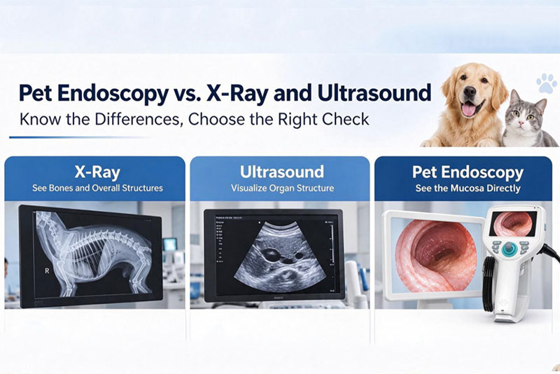
Veterinary practices often evaluate digital radiography (DR/X-ray), ultrasound and veterinary endoscopy when purchasing diagnostic systems. All three are core clinical tools, yet they differ markedly in imaging principles, clinical indications, operational costs and training requirements. No single device is universally superior; selection depends on your clinic’s caseload and service positioning.
X-rays penetrate tissue to generate 2D planar images. As a frontline outpatient and emergency screening tool, it is indicated for fractures, joint luxation, high-density foreign body obstruction, pulmonary disease, pleural effusion and intestinal pneumatosis.
Benefits: Rapid acquisition (minutes per exposure), intuitive workflow and minimal consumables, delivering low per-case operational cost. Ideal for high-volume routine and emergency triage.
Limitations: Only displays tissue contours with poor soft-tissue contrast. Small gastrointestinal mucosal defects and low-density soft foreign bodies are not detectable. Static imaging cannot visualize luminal lining or enable biopsy sampling.
Ultrasound uses reflected sound waves with no ionizing radiation. It assesses abdominal soft organs including liver, kidneys, spleen, bladder, uterus and heart, and screens for ascites, cysts, space-occupying lesions and pregnancy, with real-time visualization of organ motion.
Benefits: Radiation-free, repeatable exams. Distinguishes solid from fluid-filled lesions to characterize tissue texture. No film required; only coupling gel as a low-cost supply.
Limitations: Gastrointestinal gas severely degrades image quality during bowel distension. Cannot directly visualize mucosal ulcers/erosions, nor perform biopsy or foreign body retrieval.
The SOAIM veterinary endoscope features a high-definition camera probe. Inserted through natural orifices (oral, nasal, anal, reproductive tracts), it provides direct visualization of luminal mucosa. It is an irreplaceable intraluminal modality that X-ray and ultrasound cannot replicate.
Indications: Radiolucent soft foreign bodies (plastic bags, fabric, rubber) in cats and dogs; esophageal/gastric ulcers, polyps, chronic vomiting/diarrhoea, nasal and upper airway lesions.
Core Benefits: Diagnosis and minimally invasive treatment in one procedure. Foreign bodies can be retrieved in-situ, and suspicious lesions sampled for histopathology. It eliminates the need for laparotomy, reduces patient trauma and creates a differentiating service for clinics.
Limitations: Limited to intraluminal viewing; cannot penetrate organ walls to evaluate extraluminal pathology. Comprehensive abdominal screening still requires X-ray or ultrasound. The system has a gentle learning curve with manufacturer hands-on training. Disposable accessories include biopsy forceps and retrieval baskets.
Equipment Type | Reference Purchase Price | Learning Curve | Consumable Notes | Key Selection Criteria |
Veterinary DR (X-ray) | Entry-level: 8,500 Standard: 25,700 | Low | Minimal consumables; no single-use parts. Periodic service for detector & X-ray tube | Essential baseline unit for bone, thoracic and dense foreign body emergency screening; radiation protection mandatory |
Veterinary Ultrasound | Entry-level portable B&W: 4,300 Standard colour Doppler: 17,100 | Moderate; hands-on scanning training required | Virtually no disposables; coupling gel low-cost | Soft-tissue assessment, radiation-free. Suitable for pregnancy, ascites and hepatorenal workup; compromised by bowel gas |
Veterinary Endoscope (SOAIM) | Entry-level: 4,300 Standard HD: 11,400 | Low; manufacturer training available | Main unit reusable long-term; biopsy forceps, retrieval baskets, flushing tubes disposable | Direct intraluminal visualization, foreign body retrieval, mucosal biopsy. Requires anaesthetic monitoring; enables affordable minimally invasive services for small clinics |
DR and ultrasound are foundational imaging systems with moderate upfront cost and low running expenses, making them the first priority for most veterinary clinics. The entry-level SOAIM veterinary endoscope carries a low capital barrier and simple operation, enabling practices to launch minimally invasive services efficiently.
The durable main unit requires only limited disposable accessories. It addresses soft foreign bodies and gastrointestinal mucosal disease that cannot be diagnosed indirectly via X-ray or ultrasound, raising case value and reducing referral loss.
Key takeaway: These modalities complement, rather than replace, one another. Complex cases often require combined imaging for definitive diagnosis. A balanced equipment portfolio expands service capability and improves diagnostic confidence.
Q: When buying veterinary endoscopy, should I focus on operational complexity and staff training cost?
A: SOAIM endoscopes are designed for ease of use. Manufacturer-provided standardized practical training allows veterinarians to quickly master foreign body retrieval and biopsy to grow high-margin services.
Q: Are ongoing consumable costs high for endoscopy?
A: The main endoscope console has a long service life. Costs stem primarily from single-use accessories such as biopsy forceps and retrieval devices. Consumption scales with caseload. Compared with open laparotomy, total consumable expenditure for intraluminal minimally invasive procedures is cost-effective.
Q: For small clinics, purchase endoscopy first or X-ray/ultrasound?
A: X-ray and ultrasound are mandatory baseline screening tools and should be installed first. Add SOAIM endoscopy later if your clinic sees frequent chronic GI cases and soft foreign-body ingestions, to build differentiated minimally invasive offerings.
Q: Does endoscopy require a dedicated operating room?
A: A standalone OR is unnecessary. However, anaesthetic monitoring and oxygen delivery equipment are required for sedation or general anaesthesia. The examination area simply needs proper cleaning and disinfection. It fits small-to-medium clinics and occupies less space than open surgery.
Q: What clinical and commercial gains come from adding SOAIM endoscopy alongside existing X-ray and ultrasound?
A: X-ray and ultrasound only deliver indirect imaging findings. Endoscopy enables direct visual confirmation plus minimally invasive intervention. It lets your practice manage high-value cases including soft foreign body removal and GI biopsy, reduce referral leakage, increase revenue per case and strengthen your clinic’s technical reputation.