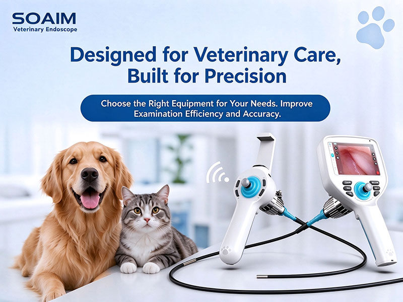
Veterinary endoscopes are essential tools for visual examination of the gastrointestinal tract, ears, and respiratory tract in dogs, cats, and other animals. Depending on the configuration, they can also support foreign body retrieval, biopsy sampling, and minimally invasive procedures. With different patient sizes, examination sites, and procedural requirements, how do you choose the right equipment?
This guide covers six key factors: insertion tube diameter, image quality, operating features, system design, maintenance and service, and overall cost.
Choose the Insertion Tube Diameter for the Animal and Examination Site
The insertion tube diameter affects access to narrow anatomical structures and can influence the internal design and available features. Consider the following factors:
Animal size and examination site: Select an appropriate outer diameter based on the species, patient size, examination site, and anatomical structure.
Procedure requirements: For visual examination alone, consider the diameter, working length, and flexibility. For biopsy or foreign body retrieval, check whether a working channel is available.
Available features: Working channel dimensions and articulation mechanisms may vary with tube diameter. The thinnest option is not necessarily the best choice for every procedure.
Image quality affects how clearly anatomical details and potential lesions can be visualized. Focus on three key factors:
Resolution: Determines how much fine detail can be captured.
Depth of field and field of view: Affect the range of distances that remain in focus and the area visible in a single view.
Illumination: Consider brightness, uniformity, and image quality during close-up examination.
Evaluate product specifications alongside actual images or a live demonstration to assess detail, color reproduction, illumination, and image stability.
Articulation and channel configuration determine which examinations and procedures the equipment can support.
Tip articulation: Check the available directions, angulation range, and ease of control.
Working channel: For biopsy or foreign body retrieval, verify the channel's inner diameter and compatibility with the required instruments.
Irrigation and suction: Confirm whether these functions and their associated ports are available for your intended procedures.
Not all veterinary endoscopes support biopsy, irrigation, or suction. Always check the specifications of the specific model.
The system design affects daily handling, image display, and mobility. Common options include the following.
These systems combine the display, image processing, and controls in a handheld unit.
Advantages: Highly integrated and convenient to operate without an external display.
Considerations: Screen size is fixed, so check device weight, battery life, and handling comfort.
Suitable for: Routine consultations, mobile examinations, and settings where standalone operation is preferred.
These systems connect to smartphones, tablets, or computers via Wi-Fi or a wired connection.
Advantages: Flexible display options and convenient image sharing for mobile examinations and teaching demonstrations.
Considerations: Check device compatibility, connection stability, and software features.
Suitable for: Mobile examinations and applications requiring flexible display options.
These systems typically include a monitor, image processor, light source, and auxiliary components.
Advantages: Larger displays and a more comprehensive system configuration.
Considerations: Require more space, are less mobile, and generally involve higher purchase and maintenance costs.
Suitable for: Dedicated examination rooms and specialized clinical settings.
Selection tip: For routine clinical work and mobile examinations, consider portable systems with integrated displays or external connectivity. Integrated systems prioritize standalone operation, while connected systems offer greater display flexibility. Choose according to your workflow and viewing requirements.
Long-term usability depends not only on the equipment itself but also on its maintenance requirements and service support.
Cleaning and disinfection: Confirm the approved cleaning and disinfection methods for the insertion tube, handle, and main unit.
Accessory availability: Check the availability of replacement insertion tubes, compatible instruments, and other accessories.
Training: Find out whether operating guidance and maintenance training are provided.
After-sales service: Review the warranty period, repair process, and availability of replacement parts.
Different components may have different cleaning and disinfection requirements. Always follow the manufacturer's instructions for use.
The purchase price alone does not reflect the full cost of veterinary endoscopy equipment. Consider both the initial investment and long-term operating expenses.
Initial purchase cost: Include the main unit, insertion tube, display terminal, and necessary accessories.
Expansion costs: Check whether additional tube diameters or configurations require a separate main unit or articulation mechanism.
Operating costs: Consider consumables, replacement insertion tubes, and routine maintenance.
Service costs: Review warranty coverage, repair charges, and spare-part availability.
Compare systems with similar specifications and features to assess their overall value. For clinics that need to cover multiple examination scenarios, a modular system compatible with different insertion tubes may help reduce duplicate equipment purchases while meeting a wider range of needs.
Choosing the right veterinary endoscope starts with identifying the target species, examination sites, required procedures, and daily workflow. Then compare equipment specifications, maintenance requirements, and total cost of ownership to find the most suitable solution.
SOAIM specializes in the development of endoscopic visualization equipment and provides product information and selection support for different veterinary applications. Contact us to learn more about suitable options for your needs.