Gastrointestinal biopsy performed using a veterinary flexible endoscope is an essential diagnostic procedure for dogs and cats with chronic vomiting, melena, or unexplained weight loss. Many new practitioners mistakenly view biopsy simply as “pinching off a piece of mucosa.” When collecting samples with a veterinary flexible endoscope, biopsy site selection, technique, and sample handling directly determine the validity of histopathology results. Improper technique can also lead to complications including hemorrhage and gastrointestinal perforation.
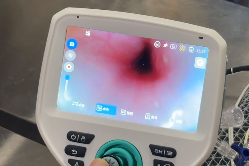
Fasting: Dogs require a 12–24 hour fast, while cats fast for 8–12 hours. This empties gastric contents and prevents visual obstruction during evaluation with the veterinary flexible endoscope.
Equipment: Perform a leak test prior to every use of the veterinary flexible endoscope. Verify tip deflection, air/water delivery, and suction function. Use disposable serrated biopsy forceps sized to match the scope’s working channel. Forceps jaws must remain closed while advancing or withdrawing through the channel to avoid scratching the working channel of the veterinary flexible endoscope.
Anesthesia: General anesthesia with endotracheal intubation and continuous patient monitoring is required to prevent aspiration.
Contraindications: Biopsy with the veterinary flexible endoscope is not recommended for patients with coagulopathies or acute gastrointestinal perforation.
When advancing the veterinary flexible endoscope, follow this core rule: advance only when the lumen is visible; withdraw if the lumen cannot be identified. A red-out view indicates the scope tip is pressed against the mucosa. Immediately retract the scope, adjust the angle, and gently insufflate air to open the lumen. Never advance the scope forcefully, as this can traumatize the mucosa.
Thoroughly examine the esophagus, stomach, and duodenum. Capture images and document the location of all lesions.
Visible lesions: Sample the margins of the lesion first. Avoid sampling only necrotic tissue from ulcer surfaces.
Chronic cases with grossly normal mucosa: Perform multiple random biopsies from the gastric fundus, body, and antrum. Collect a minimum of 6–8 adequate specimens to minimize false-negative results.
1. Manipulate the veterinary flexible endoscope to position the scope tip as perpendicular to the mucosal surface as possible.
2. Close the biopsy forceps and advance them through the working channel of the veterinary flexible endoscope until the forceps tips come into view.
3. Open the jaws and gently appose the mucosa. Gentle suction may be applied before closing the forceps. Do not apply excessive pressure.
4. Keep the jaws closed and withdraw the forceps slowly and steadily. Avoid violent tearing of the mucosa.
5. Carefully place the tissue specimen onto filter paper, flatten it, and transfer it into 10% formalin fixative. Do not compress the tissue.
6. After each sample collection, hold position for 30–60 seconds to assess for active bleeding. Minor oozing may be controlled with cold saline lavage and suction.
Do not perform biopsy with the veterinary flexible endoscope when visualization is poor or food debris obscures the field.
Advancing or retracting forceps with open jaws will scratch the working channel of the veterinary flexible endoscope and damage the scope.
Reduce the number of biopsy samples for ulcerated or neoplastic lesions; monitor closely for perforation risk.
Rolled or compressed tissue creates histologic artifacts, rendering slides uninterpretable for the pathologist.
Once biopsy sampling is complete, slowly withdraw the veterinary flexible endoscope and re-evaluate all biopsy sites for delayed bleeding. Continue postoperative monitoring and discuss potential risks with pet owners.
New operators should begin training on ex vivo tissue models. Start with straightforward exploratory cases using the veterinary flexible endoscope. Master basic skills before tackling high-risk cases such as deep ulcers and neoplasia.
> Takeaway: Gastrointestinal biopsy using the veterinary flexible endoscope depends on precise sampling, atraumatic manipulation, and standardized sample preservation, balancing diagnostic yield and patient safety.