Case | Chronic Gastritis with Gastric Mucosal Erosion in a Cat — Follow-up Gastroscopy After Medical Treatment |
Location | Shenzhen, China |
Veterinary Hospital | Wangwang Pet Hospital |
Application | Post-treatment follow-up and assessment of gastric mucosal recovery |
Configuration | SOAIM SAPro veterinary endoscopy system + Ø5.8 mm insertion tube, providing higher image resolution and a wider field of view |
Key Features | 1 MP HD real-time imaging; integrated irrigation and suction; 140° wide-angle field of view; 360° articulation; accessory channel for interchangeable instruments |
Many pet owners face a similar situation: after a period of medical treatment for gastritis, their cat stops vomiting, regains its appetite, and becomes more active. On the surface, the cat appears to have recovered.
However, improvement in clinical signs does not necessarily mean that the gastric mucosa has fully healed. In cats that previously showed gastric mucosal congestion, erosion, or other abnormalities, appetite, activity, and vomiting alone cannot directly indicate the current condition of the stomach lining.
When further assessment is clinically indicated, follow-up gastroscopy allows veterinarians to directly examine the stomach and previously affected areas. By comparing current findings with earlier endoscopic images, veterinarians can obtain additional visual information to support ongoing clinical assessment.
So, what does a veterinarian look for during follow-up gastroscopy after treatment for feline gastritis? What challenges need to be addressed during the examination? The following case from Wangwang Pet Hospital in Shenzhen provides a practical example of how veterinary gastroscopy can support post-treatment follow-up.
During the initial gastroscopic examination, the primary focus is to examine the stomach and identify and localize any abnormal findings.
After treatment, the focus changes: how have the previously identified lesions changed, and what is the current condition of the gastric mucosa?
For example, if mucosal congestion or erosion was observed during the initial examination, the veterinarian can focus on the same areas during follow-up, assessing changes in mucosal appearance, color, and surface condition.
When examination images are properly archived, the two examinations can be compared to create a continuous visual record of disease progression and recovery.
Follow-up gastroscopy is therefore more than simply “looking again.” It provides visual comparison of changes in the gastric mucosa following treatment.
Of course, not every cat with gastritis requires repeat gastroscopy. Whether a follow-up examination is appropriate, and when it should be performed, should be determined by the veterinarian based on the cat’s clinical condition, treatment history, and response to therapy.
Wangwang Pet Hospital in Shenzhen treated a cat that presented with frequent vomiting, reduced appetite, and poor overall condition. Following examination, the cat was diagnosed with chronic gastritis accompanied by gastric mucosal erosion.
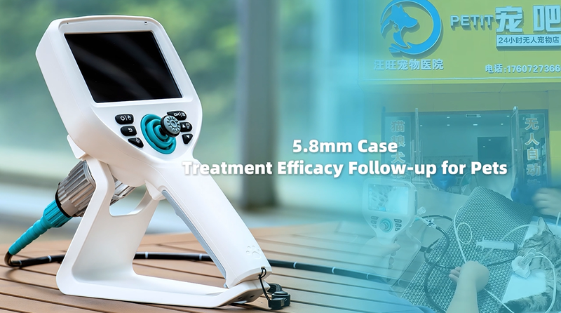
After a period of treatment, the cat showed significant clinical improvement: vomiting had largely stopped, while appetite and activity gradually returned.
However, to further assess the condition of the gastric mucosa, the veterinarian scheduled a follow-up gastroscopic examination based on the clinical findings.
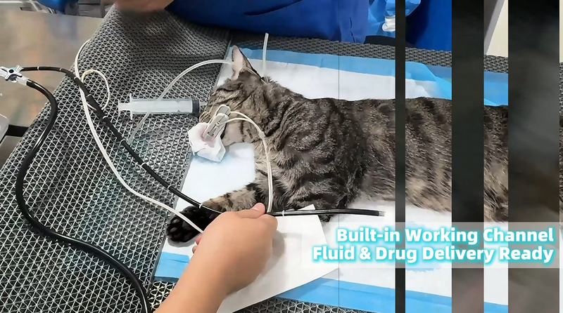
The examination was performed using the SOAIM SAPro veterinary endoscopy system, equipped with a Ø5.8 mm insertion tube to examine the stomach.
During the follow-up examination, the veterinarian first located the previously identified lesion and then assessed the surrounding gastric mucosa and overall condition of the stomach.
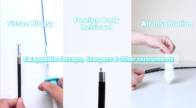
Compared with assessing clinical signs alone, gastroscopy provides direct visualization of the gastric cavity. In cases requiring comparison over time, image quality, scope maneuverability, field of view, and the ability to maintain a clear visual field all contribute to examination efficiency and the quality of documented findings.
Dr. Chen of Wangwang Pet Hospital, who has extensive clinical experience in gastrointestinal disease and endoscopic procedures in dogs and cats, commented:
“After the clinical signs of gastritis subside, the condition of the gastric mucosa may still need to be assessed. For cases that require follow-up, we pay close attention to changes between examinations. The SAPro provides clear imaging and responsive scope control, while its irrigation, suction, and image-recording capabilities are convenient for clinical follow-up.”
A follow-up gastroscopic examination involves more than simply inserting an endoscope into the stomach. Because the gastrointestinal tract of a cat provides limited working space, veterinarians need to locate previous lesions, examine the mucosa, and document their findings. This requires the imaging, maneuverability, and workflow of the endoscopy system to work together.
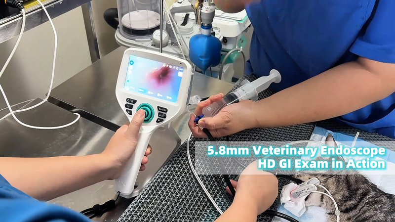
Clear visualization is fundamental to follow-up examination.
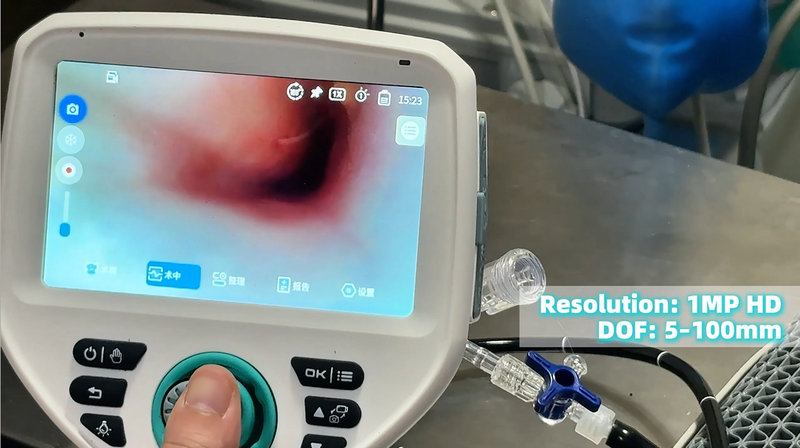
The SAPro provides 1 MP HD real-time imaging, delivering detailed views of the stomach and providing a clear visual basis for assessing mucosal color, surface characteristics, and changes in previously identified lesions.
Examination images can also be saved for future reference, making it easier to compare current findings with previous examinations during follow-up visits.
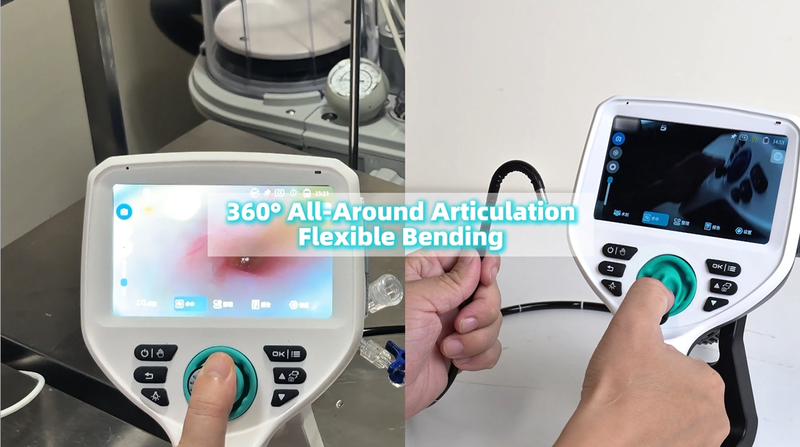
The stomach has a complex internal structure, and locating a previously identified lesion during follow-up requires precise scope control and a sufficiently wide field of view.
The SAPro provides 360° articulation and a 140° wide-angle field of view, allowing veterinarians to adjust the scope direction and viewing angle according to the examination site.
This flexibility can also improve efficiency when comparing specific areas of the gastric mucosa over time.
Gastric fluid and secretions can interfere with visualization. The SAPro integrates irrigation and suction, allowing the veterinarian to clear the field of view when necessary during the examination.
The system also supports the storage of examination images and reports, helping create a more complete clinical record that can be referenced during subsequent visits and follow-up assessments.
For this cat, the initial gastroscopy focused on identifying abnormalities in the gastric mucosa, while the follow-up examination focused on how those abnormalities had changed after treatment.
From lesion detection and diagnostic support to post-treatment follow-up and monitoring, veterinary endoscopy enables veterinarians to directly visualize the gastrointestinal tract at different stages of clinical care. Archived images can also provide useful reference material for subsequent examinations and clinical assessment.
Cats cannot tell us whether their gastric mucosa is healing. Owners can observe improvements such as less vomiting, better appetite, and increased activity, but veterinary gastroscopy allows clinicians to directly assess changes inside the stomach.
SOAIM continues to focus on veterinary endoscopy technology and product development, providing solutions for canine and feline gastroscopy, veterinary gastrointestinal endoscopy, and other clinical endoscopic applications.
Every view brings greater confidence in care.
Learn more about the SOAIM SAPro veterinary endoscopy system and solutions for gastrointestinal endoscopy in dogs and cats.
Veterinary gastroscopy allows veterinarians to directly examine the inside of the stomach, including the color, shape, and surface characteristics of the gastric mucosa, as well as abnormalities such as congestion, erosion, and ulceration.
For examinations that require close assessment of mucosal details, HD imaging provides clearer visualization of tissue features, helping veterinarians assess lesions, document findings, and compare changes before and after treatment.
The SAPro provides 1 MP HD real-time imaging to support clear visualization during canine and feline gastrointestinal endoscopy.
A Ø5.8 mm insertion tube may be suitable for certain cats and small dogs, depending on the animal’s size, the examination site, and the clinical requirements.
The appropriate insertion tube diameter should be selected by a veterinary professional based on the specific patient and procedure.
For invasive procedures, veterinary endoscopes should undergo a standardized reprocessing procedure that typically includes pre-cleaning, enzymatic cleaning, rinsing, disinfection, and final processing.
For the SAPro, the insertion tube is cleaned with a 1:200 dilution of enzymatic detergent and then disinfected using 0.55% ortho-phthalaldehyde (OPA). The standard immersion time is 5 minutes for routine cases and 10 minutes when used on animals with infectious diseases.
Specific procedures should always follow the product instructions and the veterinary hospital’s infection-control protocols.
Previous gastroscopy images can provide useful reference material during follow-up examinations. By comparing images from different examinations, veterinarians can focus on specific areas and assess changes in previously identified lesions.
Yes. Veterinarians can select examination images, annotate key findings, and add diagnostic recommendations to generate a clinical report with a single operation.
Reports can also be accessed quickly via a LAN-based QR code, making it easier to share examination records and manage clinical cases.